Saturday, October 8, 2011

Occipital Encephalocele


Encephalocele

Two major forms of dysraphism affect the skull, resulting in protrusion of tissue through a bony midline defect, called cranium bifidum. A cranial meningocele consists of a CSF-filled meningeal sac only, and a cranial encephalocele contains the sac plus cerebral cortex, cerebellum, or portions of the brainstem. Microscopic examination of the neural tissue within an encephalocele often reveals abnormalities. The cranial defect occurs most commonly in the occipital region at or below the inion, but in certain parts of the world, frontal or nasofrontal encephaloceles are more prominent. These abnormalities are one tenth as common as neural tube closure defects involving the spine. The etiology is presumed to be similar to that for anencephaly and myelomeningocele; examples of each are reported in the same family.
Infants with a cranial encephalocele are at increased risk for developing hydrocephalus due to aqueduct stenosis, Chiari malformation, or the Dandy-Walker syndrome. Examination may show a small sac with a pedunculated stalk or a large cystlike structure that may exceed the size of the cranium. The lesion may be completely covered with skin, but areas of denuded skin can occur and require urgent surgical management. Transillumination of the sac may indicate the presence of neural tissue. A plain roentgenogram of the skull and cervical spine is indicated to define the anatomy of the vertebrae. Ultrasonography is most helpful in determining the contents of the sac. MRI or CT further helps define the spectrum of the lesion. Children with a cranial meningocele generally have a good prognosis, whereas patients with an encephalocele are at risk for visual problems, microcephaly, mental retardation, and seizures. Generally, children with neural tissue within the sac and associated hydrocephalus have the poorest prognosis. Meckel-Gruber syndrome is a rare autosomal recessive condition that is characterized by an occipital encephalocele, cleft lip or palate, microcephaly, microphthalmia, abnormal genitalia, polycystic kidneys, and polydactyly. Determination of maternal serum α-fetoprotein levels and ultrasound measurement of the biparietal diameter as well as identification of the encephalocele itself may diagnose encephaloceles in utero.

Contact: Dr Deepak Dadge on face book.


Tuesday, March 1, 2011

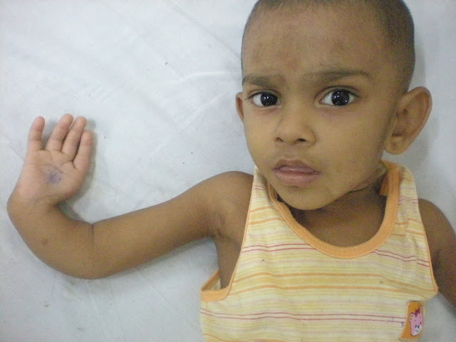
Answer: Picture no. 2


Question: Name the syndrome and congenital anomalies associated with this syndrome?

Answer: Holt-Oram syndrome. Absent thumb with congenital heart disease.


Background
Holt-Oram syndrome (heart-hand syndrome) is an autosomal dominant inherited disorder characterized by abnormalities of the upper limbs and heart (ASD, VSD). Holt and Oram first described this condition in 1960 in a 4-generation family with atrial septal defects and thumb abnormalities.

Pathophysiology
The syndrome is inherited as an autosomal dominant trait that is completely penetrant. The disease is due to mutations in the transcription factor TBX5, which is important in the development of both the heart and upper limbs.

Upper limb involvement
Although the clinical manifestations are variable, upper limb abnormalities are always present. Abnormalities may be unilateral or bilateral and asymmetric and may involve the radial, carpal, and thenar bones. Aplasia, hypoplasia, fusion, or anomalous development of these bones produces a spectrum of phenotypes, including triphalangeal or absent thumbs. Occasionally, upper limb malformation can be sufficiently severe to produce phocomelia (a malformation in which the hands are attached close to the body); this has been termed pseudothalidomide syndrome.

Cardiac involvement
Approximately 75% of patients have some cardiac abnormality. In most patients, the abnormality is either an atrial septal defect (ASD) or a ventricular septal defect(VSD), progressive atrioventricular block and atrial fibrillation.

Frequency
Holt-Oram syndrome is the most common form of heart-hand syndrome, with prevalence estimated at 0.95 cases per 100,000 total births.

Mortality/Morbidity
Structural lesions are present at birth. Prognosis depends on the severity of the cardiac lesions.
  • Significant intracardiac shunts can be associated with sudden death or the development of pulmonary hypertension and Eisenmenger syndrome.
  • The first clinical manifestation of the disease may be heart failure, cardiac arrhythmias (including heart block), or infective endocarditis.
  • Considerable physical and psychologic morbidity may be associated with limb abnormalities, particularly in severe cases.
Sex
Holt-Oram syndrome has no sexual predilection.

Age
  • A congenital disease, Holt-Oram syndrome is present at birth. Subtle limb involvement may not become clinically apparent until later in life when the cardiac symptoms of the disease manifest or when an individual has a child with a more severe presentation of the syndrome.
  • Cardiac conduction disease is progressive with aging.
  • Middle-aged individuals often present with significant atrioventricular block or atrial fibrillation.

Clinical
History
  • Patients may have a family history of cardiac and/or limb malformation.
  • Patients may present in infancy with obvious limb malformations and/or signs of cardiac failure secondary to ASD, VSD, or cardiac conduction disease.
Physical
  • Upper limb deformity
    • Always present but may be unilateral or bilateral
    • Left-sided abnormalities often more severe than right arm or hand abnormalities
    • Unequal arm lengths due to aplasia, hypoplasia, fusion, or anomalous development of the radial, carpal, and thenar bones
    • Abnormal forearm pronation and supination
    • Triphalangeal or absent thumbs
    • Possible abnormal opposition of thumb
    • Possible sloping shoulders and restriction of shoulder joint movement
    • Phocomelia
  • Cardiac involvement
    • Bradycardia
    • Irregular pulse (ectopy)
    • Irregular pulse that occurs irregularly (atrial fibrillation)
    • Wide, fixed splitting of the second heart sound
    • Pulmonary systolic flow murmur
    • Holosystolic murmur (should raise consideration for a VSD)
  • Anomalies involving any of the following are indicators that a diagnosis of Holt-Oram syndrome can be excluded:
    • Ulnar bone
    • Lower limbs
    • Kidneys
    • Eyes
    • Auditory
    • Craniofacial
    • Vertebrae (may or may not occur in Holt-Oram syndrome)

Causes
  • Molecular genetic studies reveal that the disease is caused by mutations that inactivate the transcription factor TBX5 on long arm of chromosome 12. This genetic disorder that is autosomal dominant and highly penetrant.


Congenital Malformation Syndromes Associated with Congenital Heart Disease

SYNDROME
FEATURES
CHROMOSOMAL DISORDERS
Trisomy 21 (Down syndrome)
Endocardial cushion defect, VSD, ASD
XO (Turner syndrome)
Bicuspid aortic valve, coarctation of aorta
Fragile X
Mitral valve prolapse, aortic root dilatation
Deletion 5p (cri du chat syndrome)
VSD, PDA, ASD

SYNDROME COMPLEXES

CHARGE association (coloboma, heart, atresia choanae, retardation, genital and ear anomalies)
VSD, ASD, PDA, TOF, endocardial cushion defect
DiGeorge sequence, CATCH 22 (cardiac defects, abnormal facies, thymic aplasia, cleft palate, and hypocalcemia)
Aortic arch anomalies, conotruncal anomalies
Alagille syndrome (arteriohepatic dysplasia)
Peripheral pulmonic stenosis
VATER association (vertebral, anal, tracheo esophageal, radial, and renal anomalies)
VSD, TOF, ASD, PDA
PHACE syndrome (posterior brain fossa anomalies, facial hemangiomas, arterial anomalies, cardiac anomalies and aortic coarctation, eye anomalies)
VSD, PDA, coarctation of aorta, arterial aneurysms

TERATOGENIC AGENTS

Congenital rubella
PDA, peripheral pulmonic stenosis
Fetal hydantoin syndrome
VSD, ASD, coarctation of aorta, PDA
Fetal alcohol syndrome
ASD, VSD
Fetal valproate effects
Coarctation of aorta, hypoplastic left side of heart, aortic stenosis, pulmonary atresia, VSD
Maternal phenylketonuria
VSD, ASD, PDA, coarctation of aorta
Retinoic acid embryopathy
Conotruncal anomalies

OTHERS

Apert syndrome
VSD
Autosomal dominant polycystic kidney disease
Mitral valve prolapse
Carpenter syndrome
PDA
Conradi syndrome
VSD, PDA
Crouzon disease
PDA, coarctation of aorta
Cutis laxa
Pulmonary hypertension, pulmonic stenosis
de Lange syndrome
VSD
Ellis-van Creveld syndrome
Single atrium, VSD
Holt-Oram syndrome
ASD, VSD, 1st-degree heart block
Infant of diabetic mother
Hypertrophic cardiomyopathy, VSD, conotruncal anomalies
Noonan syndrome
Pulmonic stenosis, ASD, cardiomyopathy
TAR syndrome (thrombocytopenia and absent radius)
ASD, TOF
Treacher Collins syndrome
VSD, ASD, PDA
Williams syndrome
Supravalvular aortic stenosis, peripheral pulmonic stenosis


Tuesday, February 22, 2011

Case no: 2

This patient is 3 yr male child complaining of recurrent cough and fever episode since 6 months of age. He was admitted for cough and fever two times. There is deformity of right hand as shown in the clinical picture.


Question: Name the syndrome and congenital anomalies associated with this syndrome?


Thursday, February 10, 2011

Spot diagnosis of clinical pictures


Answer of Picture 1.

1) This is ***** 3 yr old complaining of not gaining weight and bony deformity gradually increasing since birth.

Question 1: Identify the condition and give 3 differential diagnosis for the cause of this condition? 

Answer 1: This is most severe or florid form of rickets. This is picture of pt of cystinosis with florid rickets.

Differential diagnosis of florid rickets: 
1) Fanconi syndrome: Cystinosis, oculocerebrorenal syndrome (Lowe syndrome).
2) X-linked hypophosphatemic rickets.
3)  Vitamin D–dependent rickets type 1, Vitamin D–dependent rickets type 2.

Causes of Rickets
VITAMIN D DISORDERS
Nutritional vitamin D deficiency
Congenital vitamin D deficiency
Secondary vitamin D deficiency
 Malabsorption
 Increased degradation
 Decreased liver 25-hydroxylase
Vitamin D–dependent rickets type 1
Vitamin D–dependent rickets type 2
Chronic renal failure
CALCIUM DEFICIENCY
Low intake
 Diet
 Premature infants (rickets of prematurity)
Malabsorption
 Primary disease
 Dietary inhibitors of calcium absorption
PHOSPHORUS DEFICIENCY
Inadequate intake
 Premature infants (rickets of prematurity)
 Aluminum-containing antacids
RENAL LOSSES
X-linked hypophosphatemic rickets[*]
Autosomal dominant hypophosphatemic rickets[*]
Hereditary hypophosphatemic rickets with hypercalciuria
Overproduction of phosphatonin
 Tumor-induced rickets[*]
 McCune-Albright syndrome[*]
 Epidermal nevus syndrome[*]
 Neurofibromatosis[*]
Fanconi syndrome
Dent disease